Digital X-ray images
 Diagnostics
 Dental implants
 Periodontitis
 For children
Diagnostics
For children
Diagnostics
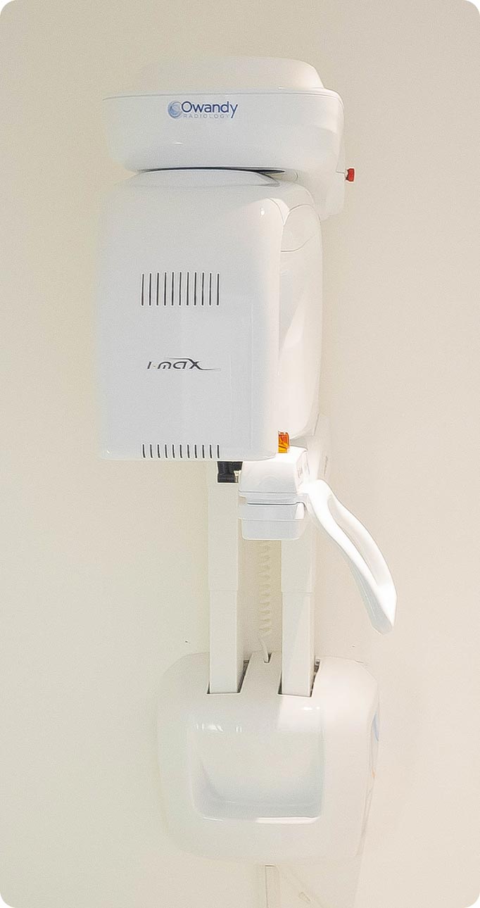
The dental panoramic is an indispensable diagnostic tool that allows the planning of the patient’s treatment. The panoramic radiological examination is characterised by its simplicity, speed and low cost.
Dental implants
The expert also needs it before a dental implant placement in order to decide whether the patient is ready for this surgical operation and whether the location of the future artificial root is ideal, for example.
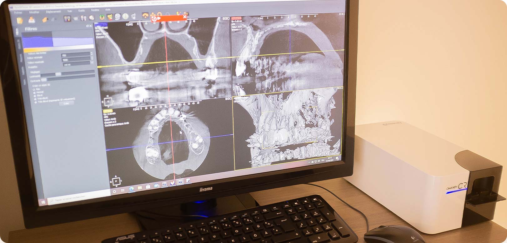
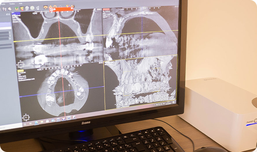
The panoramic X-ray creates an overview of the jawbone, from the crown of the teeth to the alveolar bone, including the roots and their canals.
In this way, it provides the dentist with an effective tool for determining the available space between the teeth or for checking the quality of the bone tissue.
Periodontitis
It provides valuable information about the condition of your teeth, the arrangement of your teeth and possible infections.
Infections develop in the periodontal tissue and are therefore difficult to observe without radiology. Panoramic X-rays are useful for many treatments, from orthodontics to periodontics.
For children
For children, the dental panoramic allows the visualisation of the germs of the permanent teeth, i.e. the future teeth of the adult.
This allows the evaluation of the dental age.
Cone Beam : Dental 3D scanner

Simplicity

Time saving

Low cost